
New AI tool enhances cell segmentation with gene expression data
By Charlene N. Rivera-Bonet | Waisman Science Writer
At a Glance:
- SegJointGene is a new AI software developed by the lab of Daifeng Wang, PhD, that improves cell segmentation by integrating spatial information with gene expression data.
- A unique aspect of SegJointGene is that it can output which genes are most informative for the spatial organization and segmentation of the cells.
- The new AI software outperforms current cell segmentation tools.
Understanding what a cell does starts with knowing exactly where it is. Now, a new AI-powered tool developed by researchers at the Waisman Center, University of Wisconsin–Madison, is making that task easier by combining tissue images with molecular activity data to more accurately map cell boundaries, potentially unlocking new insights into how cells work in health and disease.
The location and shape of a cell within a tissue is directly tied to its function. Learning about these cell characteristics requires a technique called cell segmentation, used to locate a cell within a tissue and draw a boundary around it, separating it from other cells. But delineating a cell becomes a challenge when cells are densely packed within a tissue, or have complex shapes, like neurons.
A new AI software, called SegJointGene, helps fine tune the process of locating and outlining cells within a tissue by combining images with the molecular information of the cells. For instance, the gene expression of a cell drives its spatial organization—the cell’s organization and shape. The tool uses gene expression data along with images of the tissue to more accurately determine the location of the cell and identify the cell’s boundaries. More accurate definition of cell boundaries, may lead to more precise insight into its function.
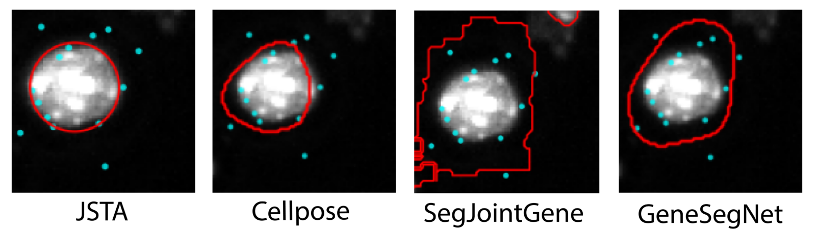
SegJointGene was developed by graduate student Haotian Ma and Daifeng Wang, PhD, H.I. Romnes Associate Professor of Biostatistics and Medical Informatics, and Computer Sciences at UW–Madison. It outperforms current tools for cell segmentation, which either rely on images alone or, when they do use gene expression, treat all genes uniformly without identifying which ones actually matter for defining cell boundaries.
The most unique aspect of the tool, Ma explains, is that it can output which genes are contributing to the spatial organization and segmentation of the cells. “This gives us two outputs. First is segmentation. Refine the segmentation. And the second is to prioritize the genes, which genes are important or play key roles to segment the cells,” Wang says.
By quantifying the amount of information each gene delivers to the cell segmentation process they can identify which genes are most important for defining that cell type’s boundaries.

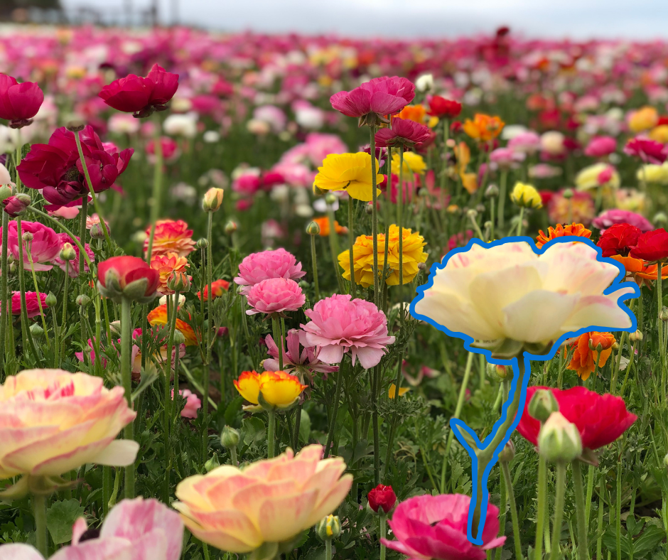
We can think of SegJointGene as identifying flowers in a diversely populated flower garden. The tool would locate and delineate each specific flower, and use their genetic composition to make sure it is accurately separating all the parts of a flower from other weeds or flowers.
“People can apply this method to any spatial data if cell segmentation is needed,” Wang says. Instead of flowers, Wang and Ma used SegJointGene to segment cells from the mouse brain and from human tonsil tissue. “But people are free to use any other omics as long as they have spatial features,” Wang says.
“The most challenging part is for the computational cost,” says Ma. Currently, analysis can take anywhere from 5-20 hours, but the researchers are working on a new version that can run faster and more efficiently. “We’re trying to apply some AI technologies to speed up processing,” he adds.
SegJointGene is free and available online for other researchers to use.